🦴 Quick Facts from Prof. Dr. Göçer
- A pathological fracture is the breaking of a bone weakened by disease due to a minor stress that would not normally cause a fracture.
- The main causes include bone metastases, primary bone tumors, benign bone lesions, and metabolic diseases such as osteoporosis.
- Preventive (prophylactic) fixation performed before a fracture occurs results in better functional outcomes and a shorter hospital stay compared to post-fracture surgery.
- In the Mirels scoring system, commonly used to assess fracture risk, a score of 8 or higher indicates the need for prophylactic surgery.
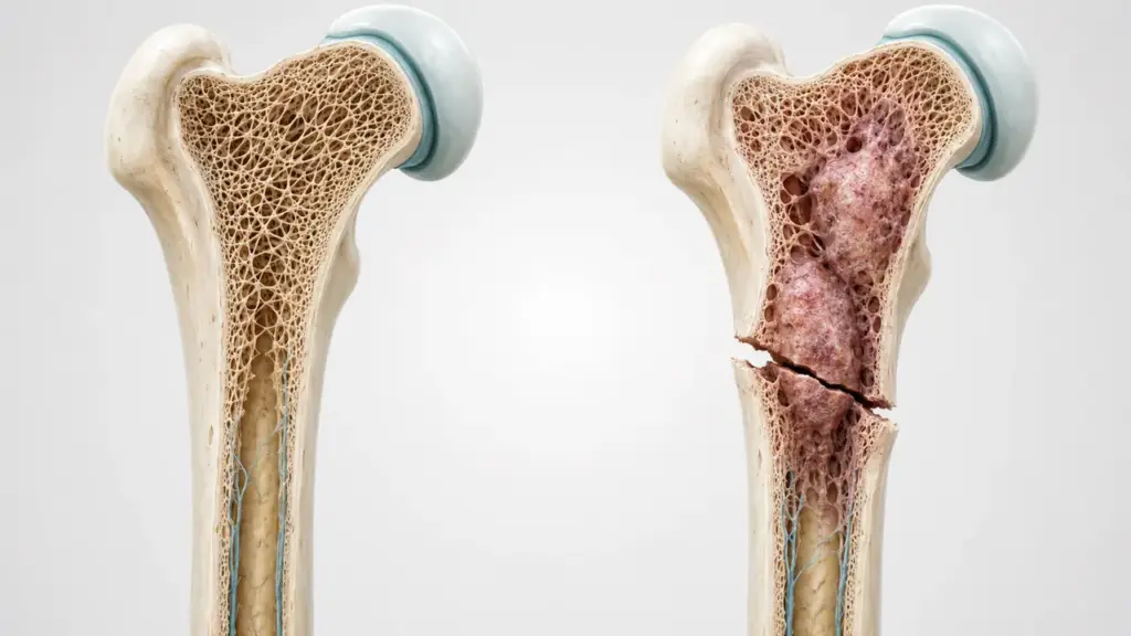
While a healthy bone may fracture only after a strong impact, some bones can fracture with much less force. A pathological fracture, is the fracture of a bone weakened by an underlying disease, either due to a force insufficient to fracture a normal bone or spontaneously. This type of fracture is often the first sign of the underlying problem in the bone.
What Is a Pathological Fracture?
A pathological fracture is a fracture that occurs against the backdrop of a disease that disrupts the bone’s structure. While a normal bone can withstand high forces, a bone weakened by a tumor, lesion, or metabolic disease loses this resistance and fractures under low-energy stress—sometimes even during everyday movements.
The most significant difference between these fractures and “normal” traumatic fractures is the presence of an underlying cause. Therefore, treatment is not limited to simply repairing the fracture; the underlying disease that caused the fracture must also be identified and managed.
What Causes Pathological Fractures? (Causes)
The main conditions leading to pathological fractures reduce bone strength in various ways. The causes of pathological fractures are generally grouped into four categories:
- Bone metastasis: The spread of cancer from another organ to the bone is one of the most common causes in adults.
- Primary bone tumors: Benign or malignant tumors originating in the bone can weaken the bone.
- Benign bone lesions: Certain benign formations, such as cysts, create cavities in the bone, increasing the risk of fracture.
- Metabolic bone diseases: Conditions such as osteoporosis reduce bone density, increasing the risk of fractures.
According to Prof. Dr. Hasan Göçer, in cases of fractures caused by unexpected trauma or those that occur spontaneously, investigating the underlying cause is just as important as repairing the fracture; because an accurate diagnosis directly determines both the treatment plan and the bone’s potential for healing.
Symptoms of Pathological Fractures
Symptoms vary depending on the location of the fracture and the underlying disease. Often, warning signs appear even before the fracture occurs. The main symptoms of pathological fractures are as follows:
- Pain before the fracture: Before the fracture, there may be progressively increasing pain in the affected area that does not subside with rest.
- Sudden pain and loss of function: Sharp pain and inability to use the affected area at the moment of the fracture.
- Swelling and deformity: Swelling, tenderness, and sometimes visible deformity at the fracture site.
- History of low-energy trauma: A fracture that occurs with a minor movement without significant trauma.
Bone pain that begins before the fracture occurs and gradually worsens may be a sign of a condition known as an “impending fracture.” An evaluation at this stage allows for preventive measures to be taken before the fracture occurs.
How Is a Pathological Fracture Diagnosed?
The goal of diagnosis is to identify both the fracture and its underlying cause. For this reason, the evaluation is more comprehensive than a standard fracture examination.
- Physical examination and medical history: The patient is asked about how the fracture occurred, previous pain, and any history of cancer.
- X-ray: It shows the fracture and areas of bone weakening (lesion, cyst, destruction).
- MRI and CT: These provide a detailed assessment of the structure and extent of the underlying lesion.
- Bone scan: This can be used to screen for other areas of involvement in the body.
- Biopsy: If the cause is unclear, a tissue sample is taken to confirm the type of lesion.
Especially in a patient without a known cancer, when a pathological fracture is detected, investigating the source is of critical importance for the treatment plan. An accurate diagnosis enables the selection of both the surgical approach and additional treatments.
Clinical Assessment by Prof. Dr. Hasan Göçer
“In my clinical practice, when I identify a lesion with a high risk of fracture in a weight-bearing bone, I prefer to perform prophylactic fixation rather than waiting for a fracture to occur. Taking this step before a fracture occurs both prevents the patient’s pain and significantly facilitates the healing process.”
Prof. Dr. Hasan Göçer — Orthopedic and Traumatology Specialist
This approach has great clinical significance. The literature reports that patients who undergo preventive fixation for an impending fracture achieve better functional outcomes and have shorter hospital stays compared to those who undergo surgery after the fracture has occurred. Therefore, not ignoring pre-fracture pain can alter the course of the process.
Treatment of Pathological Fractures
Treatment of pathological fractures pursues two goals simultaneously: stabilizing the fracture and managing the underlying disease. The decision regarding treatment is made by evaluating the location of the fracture, the underlying cause, the patient’s general condition, and the bone’s potential for healing.
Preventive (Prophylactic) Surgery
In bones at high risk of fracture but not yet fractured, stabilization is performed before a fracture occurs. This approach prevents pain and generally provides better functional outcomes than surgery performed after a fracture.
Fracture Stabilization (Fixation)
If a fracture has occurred, implants such as plates and screws or intramedullary nails are used to stabilize the bone. The appropriate method is selected based on the location; for example, intramedullary nails are often preferred for long bones.
Resection and Prosthesis
When it is necessary to remove the tumor tissue causing the fracture, the resulting void is repaired with a prosthesis or bone graft. In cases of extensive damage near a joint, a tumor prosthesis (mega prosthesis) may be used.
Prof. Dr. Göçer emphasizes that in clinical practice, the treatment method must be determined not only based on the type of fracture but also on the nature of the underlying disease. For example, in a fracture caused by metastasis, surgery is generally planned in conjunction with oncological treatment (radiotherapy, systemic therapy).
From Diagnosis to Recovery: How Does the Process Proceed?
| Stage | Time | What Happens? |
|---|---|---|
| Initial Evaluation | Day 1 | Medical history and physical examination are performed; X-rays are ordered for the fracture and the area of pain |
| Imaging & Cause Investigation | Weeks 1–2 | The underlying cause is determined using MRI/CT and, if necessary, scintigraphy |
| Biopsy (if necessary) | Depending on the situation | If the cause is unclear, the type of lesion is clarified through tissue examination |
| Surgery | Depending on the cause | The bone is stabilized through fixation, resection, or prosthesis |
| Recovery & Follow-up | Varies | Physical therapy and monitoring of the underlying condition are conducted concurrently |
Initial Evaluation and Cause Investigation: The mechanism of the fracture and any preceding pain are assessed; imaging is used to identify both the fracture and the underlying cause.
Biopsy and Surgery: If the cause is unclear, a tissue biopsy is performed. Subsequently, surgery appropriate for the type of fracture and its cause (fixation, resection, or prosthesis) is performed.
Recovery and Follow-up: Physical therapy is initiated after surgery, and the underlying condition (e.g., cancer or osteoporosis) is monitored in collaboration with the relevant departments.
Frequently Asked Questions
How is a pathological fracture treated?
Treatment aims to stabilize the fracture and manage the underlying condition. In bones at high risk for fracture, protective immobilization is used; if a fracture has occurred, fixation with a plate and screws or a nail is performed. In cases caused by a tumor, resection and prosthetics may be required. The method is determined based on the cause.
How is a comminuted fracture treated?
The goal in comminuted fractures is to realign the bone fragments into the correct position and stabilize them. Plates and screws or intramedullary nails are typically used. In comminuted fractures resulting from a pathological condition, the underlying disease must also be evaluated.
Which fracture is the most dangerous?
The danger depends on the location and cause of the fracture. Fractures in areas such as the spine and hip may be more serious due to the risk of nerve compression and loss of movement. Pathological fractures, on the other hand, are of particular importance because they indicate an underlying disease. Each fracture must be evaluated on its own merits.
Conclusion
A pathological fracture is the breaking of a bone weakened by an underlying disease due to minimal stress and is often the first sign of the underlying problem. Treatment involves both stabilizing the fracture and identifying and managing the underlying cause. Taking pre-fracture pain seriously allows for preventive measures to be taken before a fracture occurs and facilitates a better healing process.
You can begin the examination process to assess your personal situation regarding pathological fractures. Prof. Dr. Hasan Göçer evaluates each patient’s clinical presentation individually to determine the appropriate treatment options.
References
- StatPearls / NCBI Bookshelf, “Pathologic Fractures”, 2023 — https://www.ncbi.nlm.nih.gov/books/NBK559077/
- Cleveland Clinic, “Pathologic Fracture: Causes & Treatments”, 2025 — https://my.clevelandclinic.org/health/diseases/pathologic-fracture
- Frontiers in Endocrinology, “Multimodal Treatment of Bone Metastasis — A Surgical Perspective”, 2018 — https://www.frontiersin.org/journals/endocrinology/articles/10.3389/fendo.2018.00518/full